
The Optical Imaging Facility specializes in cellular and subcellular fixed or dynamic imaging and analysis of biological material. The facility provides access to powerful microscopes that enable scientists to take high-resolution pictures of stem cells. These include still images and time-lapse videos. Cells or parts of cells can be labeled with fluorescent dyes, which enable better identification of molecules and structures within cells, or to trace the fate of cells as they migrate, divide and differentiate within tissues.
-
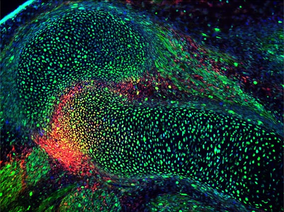
(Image by Lick Lai/ McMahon Lab) -
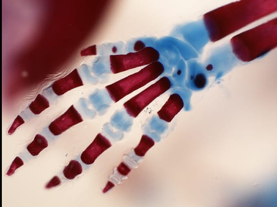
Left limb (Image courtesy of the Mariani Lab) -
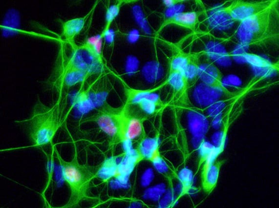
Motor neurons derived from an ALS patient (Image courtesy of the Ichida Lab) -
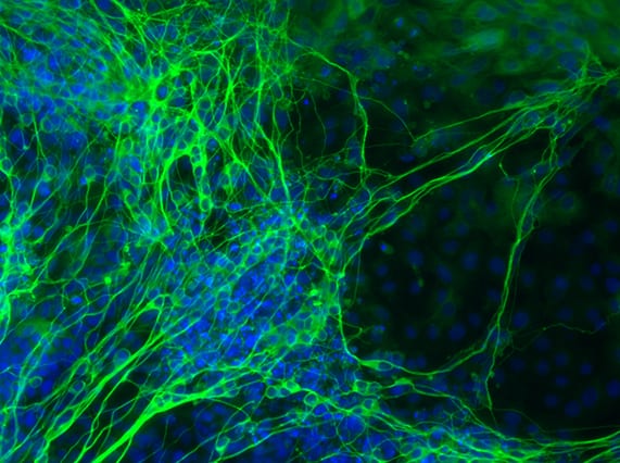
Human iPSC-derived neurons with TUJ1 staining (Image courtesy of the Ichida Lab) -

Human iPSC-derived neurons (Image courtesy of the Ichida Lab) -

Human chemical iPSC colony with NANOG-TRA-1-81-DAPI staining (Image courtesy of the Ichida Lab)